Splints In Horses
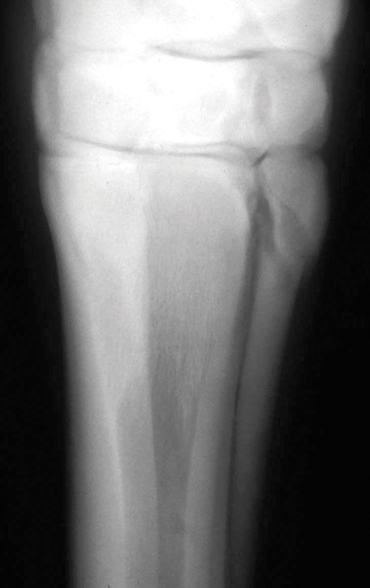
The horse has three bones in each lower leg between the knee/hock and fetlock joint. The cannon bone is the largest, and the main support for the limb. The two small splint bones, which are finger size in diameter, are long and slender and are attached to the cannon bone on each side and toward the rear. In the front legs, the splint bones are called the second and fourth metacarpals (the cannon is the third metacarpal). In the hind legs, the splint bones are called the second and fourth metatarsal bones (the cannon is the third metatarsal).
The Splint Bone
Each splint bone is held firmly against the cannon bone by a strong, thin ligament called the interosseous ligament. This forms a groove for the suspensory ligament, and also gives some protection to the deep flexor tendon that runs down the back of the leg behind the suspensory. The interosseous ligament between the splint bones and the cannon bone can sometimes be confused with the suspensory ligament because the latter is also occasionally referred to as the interosseous ligament.
The splint bones are broadest at the top, forming part of the base for the joint above it. From there on down, the splint bone tapers to a small, rounded knob. The bone ends about 2/3 of the way down the cannon, just above the fetlock joint. In an earlier time, these splint bones were probably larger leg bones. The horse’s primitive ancestors had four toes, and these bones were probably attached to the outer toes.
In the modern horse, these vestigial small bones in the front legs act as braces and support for the knee, keeping it from collapsing backward by serving as anchors for connective and supporting structures that attach to the small bone at the rear of the knee joint. This small, rounded bone (accessory carpal bone, also called the trapezium) protrudes behind the knee and acts as a brace point and fulcrum for supportive sheets of ligament that pass behind the knee from the forearm to the cannon bone.
The splint bones are not firmly attached to the cannon bone until the horse is about three years old, and sometimes as late as five years of age. Until the splint bones are completely attached, there is always the possibility of getting a splint whenever there’s too much trauma, stress, movement or rubbing between the bones. Lameness from splints is most common in two and three year olds in hard training, but has also been known to occur in horses as old as four or five that are worked exceptionally hard or have extra stress on these bones due to poor conformation.
Gary Baxter, VMD, MS, Hospital Director at University of Georgia in Athens, Georgia, says that most of what are called true splints—inflammation and subsequent bone growth due to tearing of the ligament attaching the splint bone to the cannon bone—occur in younger horses. “In an older horse with swelling in that region and lameness, you’d be more apt to suspect some external trauma,” he says.
Traditionally, it was thought that the splint bone’s primary function was weight-bearing support for the knee, alongside the cannon bone. A recent study looked at how the head of the inside splint bone interacts with the carpus (knee joint), assessing the rotational and compressive stability of the carpus. “Essentially, we found that when the top 20% of the medial splint bone was removed, it did not really affect the stability of the carpus when downward pressure was applied, but it made the carpus very unstable when the carpus was twisted. The stability was only affected when you remove the top 20% of the bone, where all the ligaments attach to the splint bone,” says Baxter.
“The very top part of the splint bone appeared to be more important to rotational stability of the carpus than it was to weight-bearing and downward compression. The head of the splint bone is essential for support of the knee during twisting actions of the limb,” he explains.
“We are not sure whether this has anything to do with the development of splints, but whenever the limb takes weight, the splint bones are pushed toward the back of the leg and probably put pressure on the interosseous ligament. Splints would be related to the movement of the splint bone and the amount of movement is probably related to the amount of work the carpus is doing. The harder the work, the more likely the interosseous ligament will tear,” he says.
Splints
 Splint bones are most familiar to horsemen in their role of creating “splints”—painful swellings that may or may not become bony enlargements. Mild irritations may cause heat and swelling, mild lameness, and then resolve. More severe cases generally produce new bone growth, and a permanent lump. After the inflammation subsides, the swelling becomes smaller and firmer. In early stages, most of the enlargement is due to inflammation, but once the area heals, all that remains is the smaller bone formation.
“From a clinical perspective, splints are seen most often in younger horses, especially when they start in training,” says Baxter. Young horses with poor front limb conformation (more stress on one side or the other) may disrupt the interosseous ligament before it stabilizes, especially if the horse is overworked.
Subsequent bony enlargements may vary from pea-size to as large as a chicken egg. A young horse in training may “pop a splint” with sudden swelling, inflammation and lameness—or may develop a bony enlargement without any history of lameness. The bony lump is usually just a blemish after the inflammatory stage is past.
“Splints are definitely more common on the inside splint bone, and many of these are fairly proximal (close to the knee). The typical location is often about 6 to 7 centimeters (2.5 to 3 inches) below the knee,” he says. Due to the shape and angle of the splint bone, the top and inside of the inner splint bone take more weight and stress than does the outer splint bone. The more weight applied to the top of the inner splint bone - as from offset cannons, often called “bench knees,” with the cannons too far to the outside rather than located directly under the knee joint - the more risk for splints. The inner splint is also more prone to injury because the inside of the leg usually takes more weight than the outside of the limb.
“People talk about the tearing of the interosseous ligament, which attaches the splint bone to the lining of the cannon bone, and I think this can be confused with the true interosseous ligament, which is the suspensory,” says Baxter.
“The suspensory ligament is between the splint bones attached to the back of the cannon bone, versus the much smaller interosseous ligament between the axial border of the splint bone and the back of the cannon bone. What most people refer to when they talk about splints is the actual tearing of that small ligament. Depending on the severity, this may involve just a little bit of local tearing—which can be resolved/healed by icing and wrapping and taking the horse out of work. In these instances, you usually don’t get any bony growth,” Baxter explains.
Bones are covered with a thin membrane (the periosteum), which protects and nourishes the bone, serves as an anchor for tendons and ligaments, and contains a layer of bone-forming cells. These cells play a role in growth and development of young bones, and in the repair of broken bones. The bone-forming cells in the periosteum may be stimulated to form new bone if injured. Inflammation and increased blood supply to the injured area can trigger new growth. This can happen anywhere along the juncture between the cannon bone and splint bone if the ligament attachment is pulled/torn.
“Whether or not a bony growth develops is often related to the severity of the injury,” says Baxter. This type of injury is generally due to excessive stress and strain on the structures, as when a young horse is being worked too hard or has poor leg conformation that puts extra strain on the inner splint bone.
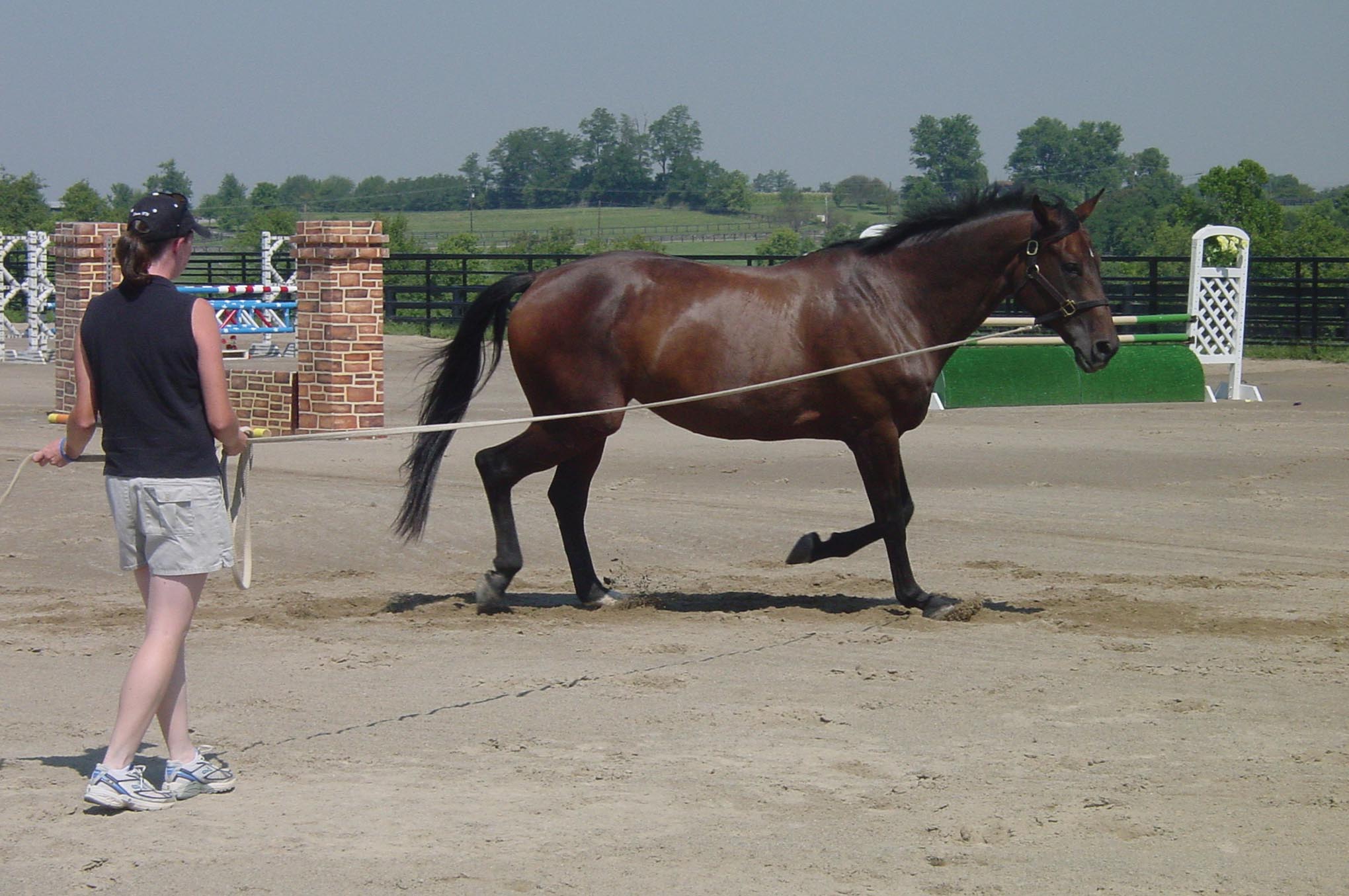
“A horse may also develop a splint from external trauma, as when a horse interferes and hits himself with the opposite foot. This may or may not be due to poor conformation.” If he strikes the splint bone, this may create injury and inflammation. Horses that are splay-footed (toe-out conformation) tend to swing the feet inward when they are in motion, and often interfere, as will some horses that are improperly shod—with feet out of balance. Exercising a young horse in small circles, such as lunging, may also contribute to splints. Not only is the horse apt to strike himself, but circling also puts more stress on the inside of the legs.
Splints may be due to immaturity and hard work. If a trainer overdoes the work, there may be tearing of the interosseous ligaments. Anything that excessively loads that inner splint bone can contribute to the formation of splints, and this includes poor conformation of the front legs.
Splints that occur on the hind legs are usually due to trauma, rather than from concussion and strain, and are much less common than splints on the front legs. Front legs bear more weight and must withstand more stress. When splints do occur on the hind legs from overwork, they are usually on the outside of the leg because the outside splint bone bears more weight and stress—with more potential for movement—which is just the opposite of what happens in the front legs.
Lameness
If the splint is acute, there will be heat, pain and swelling over the affected area. “Any pain on palpation, on the inside aspect of that inside splint bone area on a young horse, should make us suspicious of a splint. That would be our first thought, regarding the cause of lameness,” says Baxter. The horse may walk sound, but will likely be lame on that leg when trotted, especially on firm ground.
“Sometimes the swelling can become very large and contribute to chronic lameness. The question is always asked if the lameness is due to how large the bony enlargement is, or which way it grows. This can be problematic. If it grows inward, the bony enlargement may cause irritation to the main suspensory ligament or the tendons in that area. Depending on which direction the bony enlargement goes, it might cause some lameness by impinging on some of the other structures, or just from the bony lump itself,” he explains.
Many splints resolve with time and rest, without treatment. The inflammation, swelling and lameness disappear, and the horse may or may not have a bony lump in that area. The length of time it takes for the horse to become sound again will depend on the type and severity of the splint. It may be two weeks, or two months—or longer. Healing can be hastened if the inflammation can be reduced in the early stages.
Treatment
 For a bony growth that is impinging on moving parts, part of the lump can be shaved off surgically. “Even if it is right up against the knee, you can’t take out all of the splint bone because it is fairly supportive of the knee joint, but a veterinarian can surgically remove part of it with a chisel and mallet to take off the bony growth and smooth it up, to make the horse sound. This is an option for cases where the boney lump is very large, for whatever reason,” says Baxter.
“The majority of splints don’t need surgery, and the prognosis is usually very good. With surgical removal, sometimes you get recurrence of bony growth because you’ve disturbed, irritated and stimulated the bone-forming cells in the periosteum again. This causes it to lay down more layers of bone. Usually you don’t get as much bony regrowth, however, as the original lump. So even if you get a little bit of regrowth, the surgery is usually still an improvement,” explains Baxter.
Generally, however, splints are not a complicated problem. “It tends to be more of an issue for owners with show horses. Some owners want to remove a splint, just to minimize the cosmetic blemish. With the majority of splints I’ve removed, it was because they were causing lameness, or it was for cosmetic reasons. Either way, you can improve them, but for a show horse you also have to worry about the hair changing color if you do surgery,” he says.
“Any splint can result in a bony lump, or at least a temporary thickening of the periosteum and the ligament. Even if it’s not a bony enlargement, it might be a soft-tissue fibrous enlargement in that area. This kind of enlargement may tend to resolve with time, but if it is re-injured it could revert back to inflammation and a bony enlargement as well.” The best thing to do if you want to figure out whether it’s bony enlargement or just soft tissue swelling is to radiograph or ultrasound the area. Then you can decide what you might do for treatment,” explains Baxter.
“Repetitive injury and inflammation is when you end up with a bony enlargement. There are many horses that have bony enlargements on the proximal, medial splint bone (under the knee, on the inside of the leg), but if these chronic splints don’t bother the horse, they really don’t need treatment. The horse may have been a little lame at first or had some inflammation and then it quiets down and doesn’t cause any problems. So, when you see a bump on the inside of the leg, this does not necessarily mean it’s the cause of a lameness if the horse is lame on that leg.
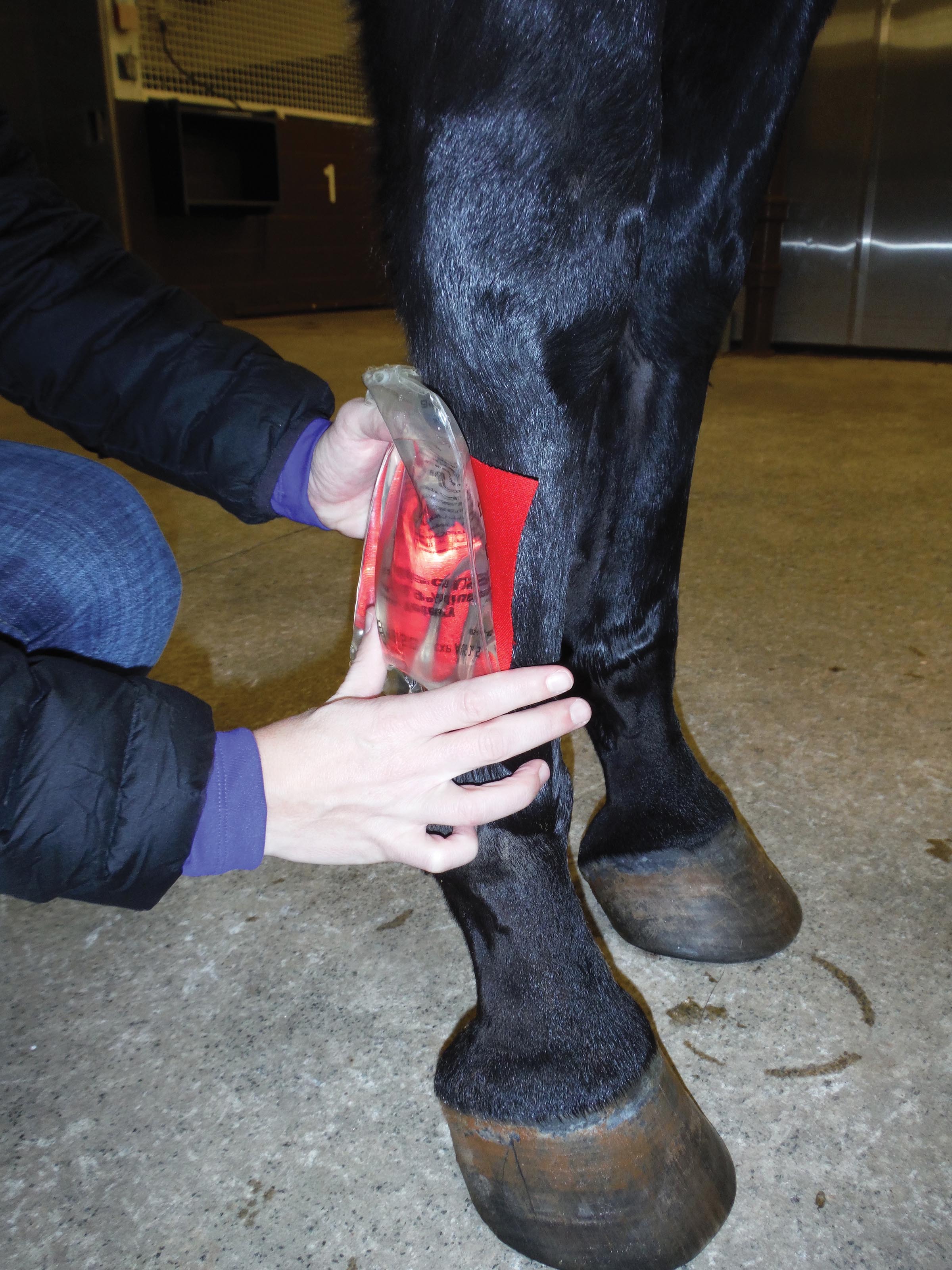
“First aid treatment on most of these, when the horse first goes lame and has heat and swelling in that area, is to decrease the inflammation. Many people recommend cold hosing or ice on the area and/or non-steroidal anti-inflammatory therapy and wrapping the leg. This is a great use for topical anti-inflammatories like Surpass or diclofenac liposome cream. Historically, we used DMSO and glycerin in a sweat, and I think this type of counter-pressure with a good snug wrap is helpful. The more you can decrease the persistent inflammatory response, the less likely the injury will result in bony enlargement,” he says. The most important thing is to reduce the inflammation.
“For chronic splints—bony lumps that still have some inflammation—some people have tried shock wave treatment to try to knock down the inflammation in the ligament itself, which could be beneficial. This type of treatment is controversial, however. Some people feel that the shock waves actually stimulate the periosteum, which could result in more bone growth,”
he explains.
Some people try counter-irritation in the acute stage. “This consists of local injection of a small dose of steroid (triamcinolone) along the ligament to really knock out the inflammation. This treatment is also controversial because the steroid may contribute to bony growth. But the whole idea is to locally treat the inflammation. You don’t want to use repeated injections of steroid because this is usually counterproductive,” he says.
“With these types of treatments, a person usually has to consider them on a case by case basis, and just select the ones that might possibly benefit,” says Baxter. Splint swelling and lameness will generally heal just fine with time, rest, and compression, but some people want to treat them more aggressively so the horse can get over the lameness and back into training more quickly.
“Horse people may use things like therapeutic ultrasound, cold laser therapy, etc., to try to reduce inflammation, but I haven’t used these treatments very much,” says Baxter. Cold therapy, topical anti-inflammatory medication, and rest are generally very adequate and effective.
 Prevention
Some people try different types of shoeing to help take stress off that part of the leg, but there’s not a lot you can do with the foot really help keep splints from occurring. “About all you can do is try to keep the feet as well balanced as possible. If horses have bench knees (with extra stress on the inner splint bone), you can’t really correct that with shoeing,” says Baxter. If you do too much “correction” you are working against the way the horse’s legs are structured and may create more stress.
“The best way to prevent splints is to take it slow with young horses in training, and not be in too big a hurry,” he says. The most common cause of splints is pushing a young horse too fast, too soon, especially on hard surfaces.
