
Knee Problems
Posted by Speedhorse on 03/29/2021
Lameness is a serious issue, particularly for an active equine athlete. Even the slightest amount of pain can impair performance, especially where seconds matter as in Quarter Horse racing. Most equine lameness in the front legs – up to 90% – is attributable to problems in the lower limb from the fetlock down. That said, a variety of injuries can occur within the knee (carpal) joints and surrounding soft tissues.
Conformational Issues:
Angular Limb Deformity
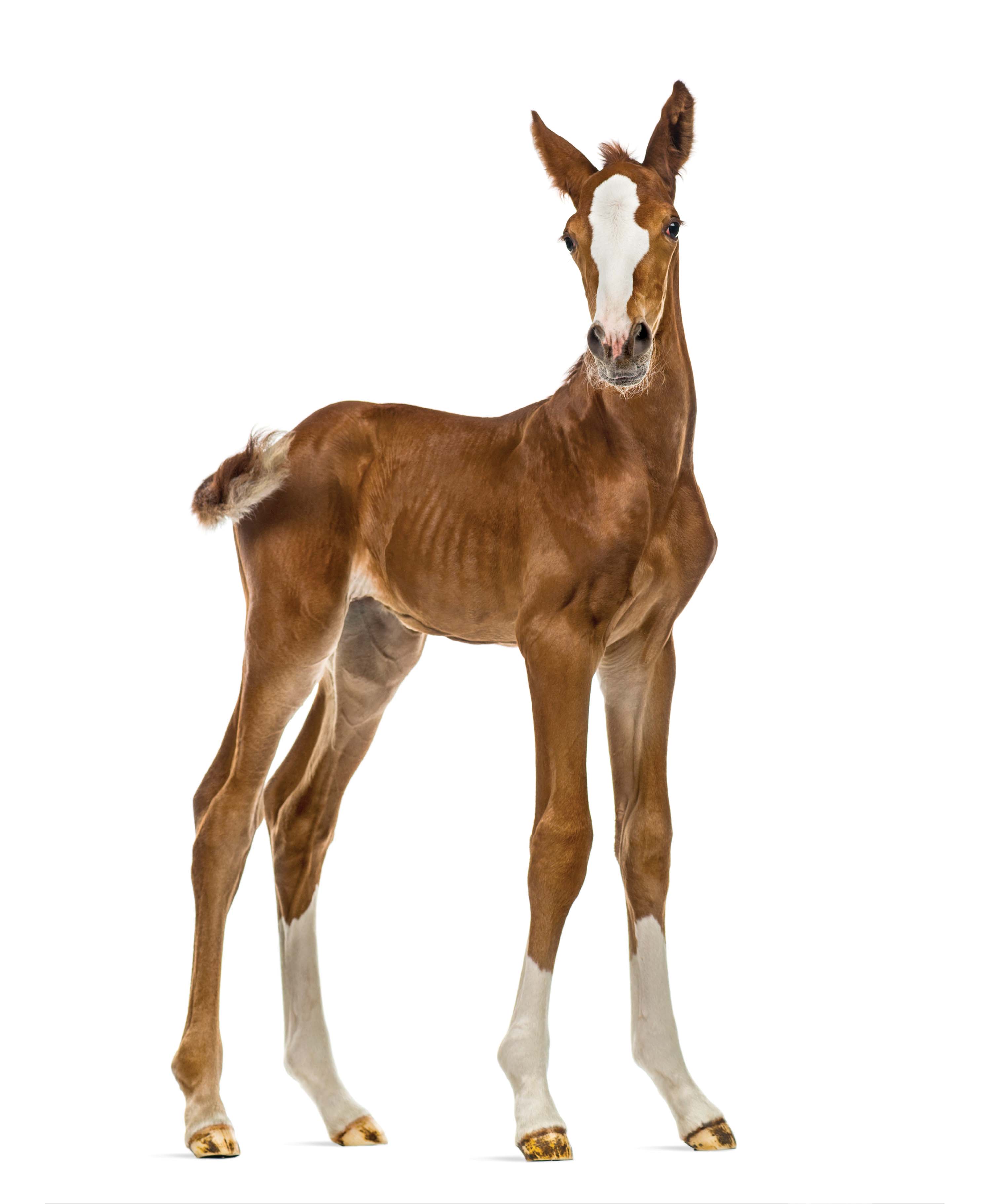 Structural stability is important to minimize stresses on equine joints. Monitoring of conformation begins at birth. A foal’s limbs aren’t always straight and perfect at the beginning. As many as 13% of foals don’t have straight limbs within the first 10 days. With growth, most spontaneously correct. Those that don’t achieve conformationally correct alignment in one or both limbs are referred to as having an angular limb deformity (ALD). While the incidence of an ALD is higher in foals with narrow chests, an ALD may occur from nutritional imbalances, rapid growth, or excess exercise or trauma, especially at a young age when the skeletal system is still developing.
Angulation of a limb outward from the knee is called carpus valgus; angulation inward (usually from the fetlock or below) is referred to as a varus ALD.
A limb experiences uneven joint loading from the carpus down through the fetlock, pastern and foot when the limb doesn’t sit straight beneath a horse’s center of mass. Ultimately this may lead to premature development of osteoarthritis and other soft tissue problems. Not all ALDs require surgical intervention, but surgery is the best option in moderate and severe cases. Initially in the first few months of life, appropriate hoof trimming and restricted exercise may help to improve the abnormal angulation.
Surgical strategies for the carpus are timed relative to status of the growth plate of the lower forearm (distal radial physis) in advance of a rapid growth phase and growth plate closure. The best chance of correction is possible if carpal surgery is performed prior to 3-4 months of age. Correction potential is significantly less successful as a foal approaches and/or passes six months of age. Early and successful correction of an angular limb deformity has no negative effects on long-term athletic performance.
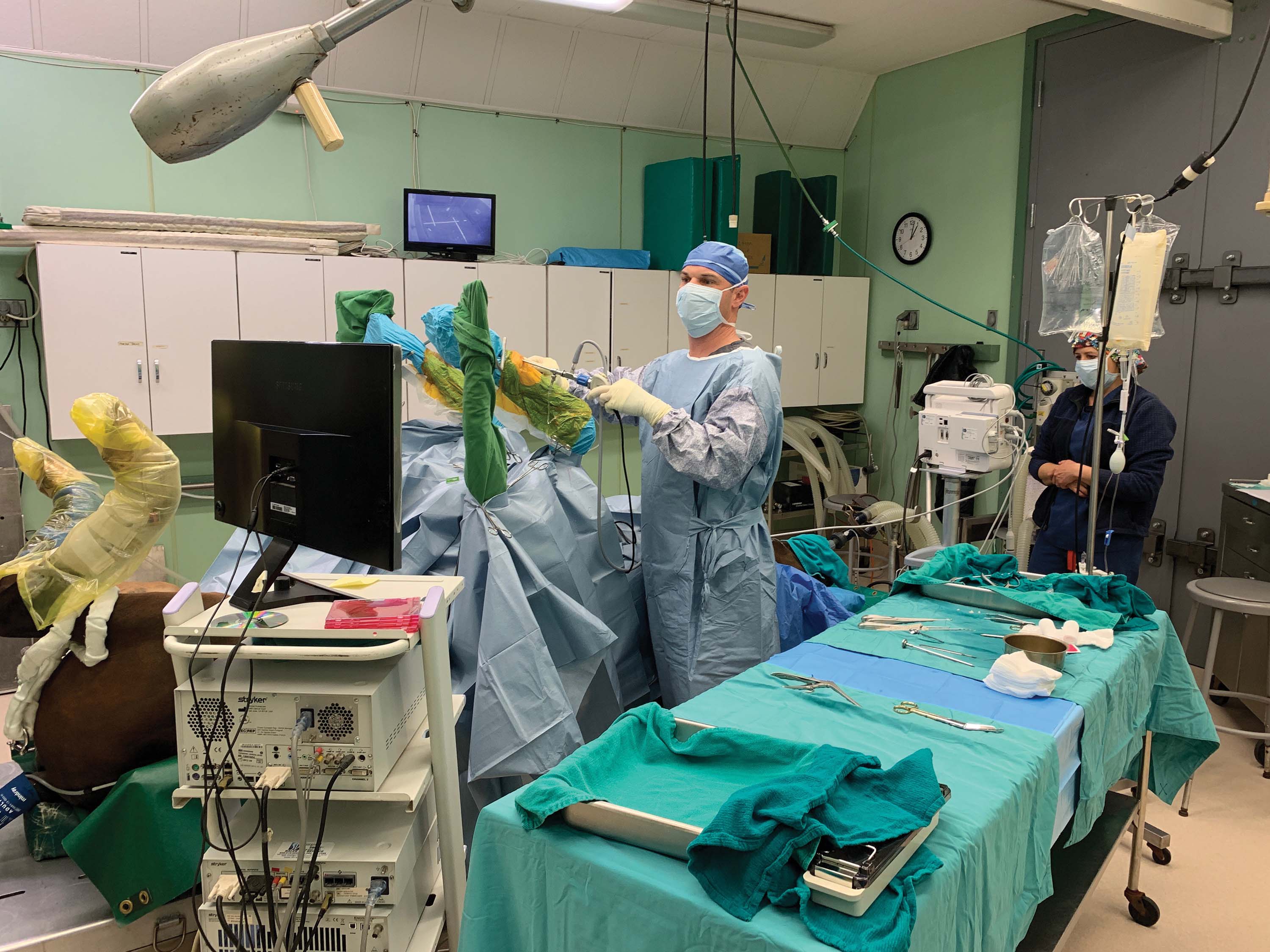 The objective of ALD surgery is to either accelerate or retard growth along the growth plate. The periosteum (nutrient-supplying tissue sheath overlying the bone) of the distal radius is stripped back to
allow the other side of the knee to “catch up.” More significant conformational deviations might need growth retarded on the convex side of the limb through transphyseal bridging that inserts screws and wires and removes them once the deviation has corrected itself. If surgical correction is not done before long bone development has passed the desired age, then it is possible to remove a wedge of bone (ostectomy) to help correct limb alignment. Close monitoring with frequent checkups and radiography are important to evaluate treatment outcome.
Additional Conformational
Issues of the Knee
Additional conformation issues occur in and around the carpus. “In-at-the-knee,” “bench knees,” and “calf knees” (hyper-extension of the back-in-the-knee conformation) potentially contribute to various carpal problems later in life. Bench knees and “off-set” knees can result in trauma to the medial splint bone from interference injury.
Carpal joint arthritis doesn’t tend to develop in horses with “over-at-the-knee” conformation since carpal joints normally flex. However, if a horse is fully weight bearing as the knee flexes prematurely, injury can result -- sudden increase of tension on the superficial digital flexion tendon potentially stretches or tears tendon fibers, resulting in a bowed tendon. In
severe cases, the carpal canal becomes inflamed, similar to carpal tunnel
syndrome in humans.
Individuals that are “over-at-the-knee” at an early age likely remain that way, whereas those that are ‘back-in-the-knee’ have more of a chance to improve between weanling age and three years old. Back-in-the-knee conformation puts undue pressure on the front margins of the carpal bones, with the potential to cause fractures or degenerative joint disease.
Many of these variations in knee conformation are genetically linked, so selection of both mare and stallion is important to maximize optimum structural features.
Acquired Knee Problems
Hygroma
Swelling over the front of the knee may be present due to development of a hygroma, which is an acquired bursa, i.e. a pocket with a secretory lining that produces serum-like fluid. Hygromas typically result from trauma, such as experienced through kicks, falls, other impacts such as banging a knee on the stall or trailer walls, or from limb entrapment in panels, fencing, or feeders.
Most often, a hygroma is a cosmetic blemish rarely affecting soundness, and it rarely becomes infected. Drainage of the hygroma, along with infusion of corticosteroids and compression bandaging, can be successful except when a hygroma connects with underlying tendons or joints that continually secrete fluid, resulting in a likelihood of recurrence. Cases of reduced range-of-motion of the knee usually have more to do with underlying osteoarthritis, fibrous joint capsular restriction, or previous carpal injury and/or surgery rather than the presence of a hygroma.
Chip Fractures
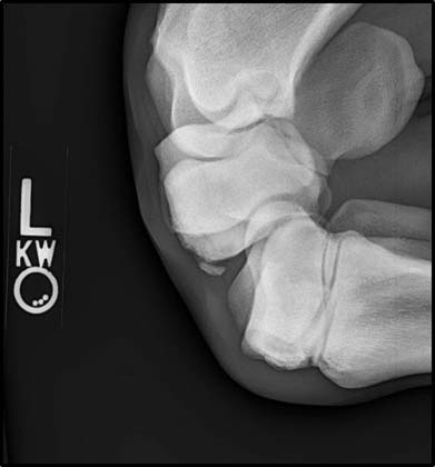 Chip fractures within the carpus are not uncommon in racehorses. Osteochondral (OC) fragments in the carpus may develop from a single yet substantial traumatic event or from repetitive trauma that exceeds bone’s capacity for adaptive remodeling. About 15% of horses have chip fractures in their joints incurred from frolic and competitive play in the field, even before training begins. Radiography can detect these before a horse enters into active training.
Horses with correct conformation that are conditioned appropriately and/or asked to perform jobs with less arduous physical impact are better protected from developing OC fragmentation. In contrast, cyclic loading, fatigue and hyperextension – activities such as racing, jumping, or sports with sharp turns – are known to precipitate OC fragmentation. Fragmentation and associated debris in the joint then lead to an irregular cartilage surface and inflammation of the synovial membrane (synovitis), with a risk of osteoarthritis. In many cases, it is best to remove carpal osteochondral fragments with surgery. Large fragments, like slab fractures, or those surrounded by scar tissue are often best left alone although some may require stabilization through arthroscopic surgery to reduce and compress the fragments.
Acute Carpitis
Acute injury to any of the multiple joints within the carpus (carpitis) can set off a cascade of inflammatory events. Radiographic and ultrasound imaging are excellent diagnostic tools to determine the extent of injury to bone and soft tissues. Rapid intervention helps to reduce internal damage within the joint. Treatment with cold therapy, systemic and local non-steroidal anti-inflammatory drugs (NSAIDs), and rest help manage acute inflammation. In some cases, arthroscopic surgery assesses the cartilage interface as well as soft tissue structures and makes it possible to flush the injured joint to reduce concentrations of inflammatory enzymes.
Soft Tissue Injury
Not all injuries within the carpus are related to internal joint or bone problems. Soft tissue structures within and around the carpus can experience injury: the superficial digital flexor tendon, the carpal canal and digital sheath, the proximal suspensory ligaments, and the intercarpal ligaments that bind the bones of the carpal joints to one another. Soft tissue injury is best identified with diagnostic ultrasound and/or MRI imaging.
Carpal Osteoarthritis (OA)
Osteoarthritis (OA) is a progressive, degenerative condition. The most effective efforts at management of OA are aimed at modification of the disease process and helping the horse deal with the symptoms of debilitation. This is usually achieved using anti-inflammatory medications, and systemic and intra-articular (IA) administration.
The horse’s carpus plays an important role in locomotion and athletic prowess. It is also susceptible to injury in a myriad of ways ranging from conformational abnormalities to repetitive-use injury to traumatic impact. Ideally, any inflammatory condition within the carpal area should be recognized and addressed as early as possible with
your veterinarian’s help to waylay the
onset of osteoarthritis.
Categories: Health